
 James
James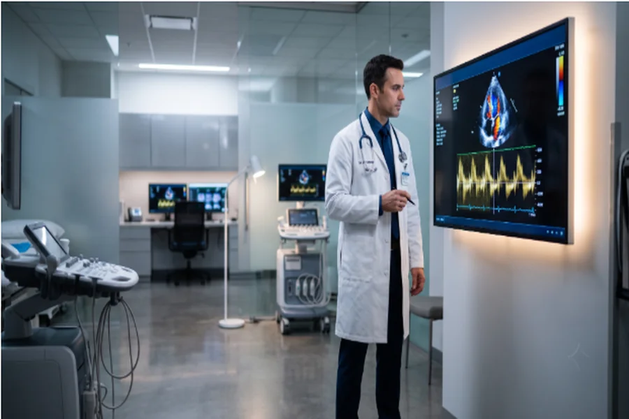
A side impact does something a front impact does not. There is no engine block, no crumple zone of any depth, and no distance between the striking vehicle and the person inside. A door panel is a few inches of sheet metal, and energy that would normally be absorbed over three feet of structure arrives almost directly at the chest.
That is why side impacts produce a category of injury that often does not appear in the initial paperwork. The chest wall can look unremarkable. The person walks out of the emergency department. Weeks later, an echocardiogram ordered for unexplained fatigue or shortness of breath shows a segment of heart muscle that is not contracting the way the segments around it are.
Why the Point of Impact Changes the Injury Pattern
Occupant protection is built around frontal collisions because that is where the available space is. Side structures have to be thin enough to fit a door, which leaves the torso close to the point of intrusion.
Force transfers through the chest wall to the structures behind it. Ribs may or may not fracture. The heart, sitting immediately behind the sternum and the left chest wall, absorbs a share of that energy whether or not anything visible breaks.
Intersections Are Where This Happens
Side impacts are overwhelmingly an intersection event. Crossing traffic, left turns across a lane, and vehicles entering from a stop are the geometries that put one bumper against another vehicle’s door.
Intersection design therefore matters more here than open-road speed. Reviews of specific problem locations, such as this look at Elizabethtown’s most dangerous intersections, tend to identify the same recurring features: heavy turning volume, obstructed sightlines, and short signal phases.
What Blunt Cardiac Injury Actually Is
Blunt cardiac injury covers a spectrum, from minor bruising of the heart muscle to severe and life-threatening damage. The clinical literature is candid about the difficulty of catching it. According to the StatPearls review on blunt cardiac injury published on the National Library of Medicine’s Bookshelf, diagnosis is challenging because there are no standardized diagnostic criteria and no single definitive test.
Evaluation relies on a combination of physical examination, electrocardiogram, cardiac biomarker monitoring such as troponin, and echocardiography where indicated. Many patients present without symptoms at first.
Reading Hypokinesis on the Study
Hypokinesis is the term for a region of the heart wall that moves less than it should. On an echocardiogram, the reading cardiologist assesses how each segment of the ventricle thickens and moves through the cardiac cycle. A bruised segment moves sluggishly while healthy tissue beside it contracts normally.
That regional pattern is the finding. It is not a general statement that the heart is weak, and not something a patient can feel. It appears only when someone orders the study and looks for it.
A Normal First ECG Is Not an All Clear
This is the part that most often creates a gap in the record. The same review notes that normal ECG and troponin results do not exclude all blunt cardiac injury, because some injuries have a delayed presentation. Where the initial twelve-lead ECG is normal, a follow-up ECG after four to six hours is recommended along with cardiac biomarker levels, and injury may take up to forty-eight hours to manifest.
A patient discharged after a single normal ECG has not necessarily been cleared. They have been screened at one point in time.
Findings That Do Not Always Resolve
The assumption that a bruise heals and the story ends is complicated by the follow-up data. A prospective study of patients with blunt thoracic trauma tracked them at three and twelve months. Among those with myocardial contusion and wall motion abnormalities, ten of seventeen still had wall motion abnormalities at three months, and four of seventeen still had them at twelve months. That is a minority, but not a negligible one, and it is the difference between a resolved event and a documented long-term change.
Why the Record Gets Thin Where It Matters Most
The sequence that produces this injury also produces a weak paper trail. The initial visit records chest wall contusion and normal vitals. The patient goes home. Symptoms developing over the following days get attributed to soreness, stress, or poor sleep. By the time an echocardiogram is ordered, weeks have passed with no documentation connecting the finding back to the collision.
Kentucky’s no-fault benefits are designed to fund exactly this kind of follow-up. KRS 304.39-020 provides basic reparation benefits of up to $10,000 per injured person for medical expenses and work loss, payable without any determination of fault, so a cardiology referral does not have to wait on an insurance dispute.
Timing rules matter too. Under KRS 304.39-230, where reparation benefits have been paid, an action for further benefits may be commenced no later than two years after the last payment, a date that does not always match the date of the crash.
What Belongs in the File
For anyone tracking a cardiac finding after a side impact, four documents carry the record: the emergency department note describing chest wall contact, the serial ECG and troponin results rather than only the first set, the echocardiogram report with its segment-by-segment wall motion description, and any repeat study showing whether the hypokinesis persisted. Together they turn an isolated imaging finding into a traceable history.
